hero image

Digital PCR assays for ovarian cancer gene variants

Order ready-to-use dPCR assays

Revolutionizing ovarian cancer research with precision dPCR assays

Explore ovarian cancer related dPCR assays by gene

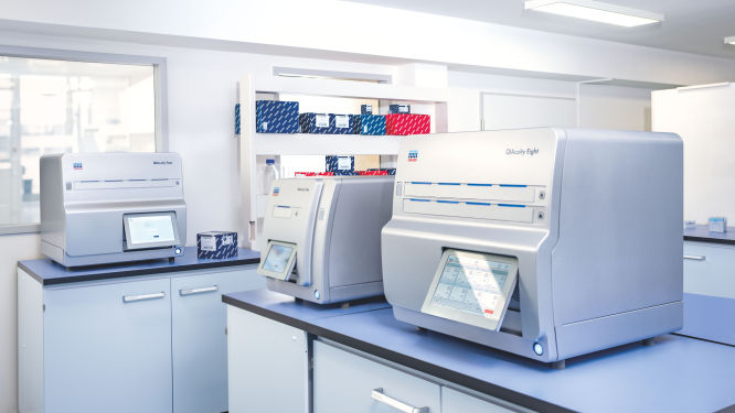
Discover the QIAcuity family of dPCR instruments

Transform your research capabilities with QIAcuity digital PCR
QIAcuity is a fully automated digital PCR system that combines precision, efficiency and ease of use. Experience unparalleled accuracy and save resources with high-throughput multiplexing, allowing for the simultaneous detection of up to five genetic targets. A seamless transition from existing qPCR workflows ensures minimal disruption while significantly enhancing data quality and throughput.
Streamline your clinical PCR workflows with QIAcuityDx
QIAcuityDx is tailored for IVD applications. This fully automated system enhances diagnostic precision and operational efficiency by reducing hands-on time and ensuring accurate detection and quantification of important genetic variations. Easily develop your own assay menu* by using QIAcuityDx utility mode and IVD medical device consumables, reagents and software.

*In compliance with the FDA ‘Medical Devices; Laboratory Developed Tests’ final rule, May 6, 2024 and European Union regulation requirements on ‘In-House Assays’ (Regulation (EU) 2017/746 -IVDR- Art. 5(5))

Frequently asked questions

How do dPCR LNA Mutation Assays benefit cancer researchers?
dPCR LNA Mutation Assays offer significant advantages to cancer researchers working on precise and sensitive mutation detection. These assays are specifically designed for use with the QIAcuity Digital PCR System and are enhanced with Locked Nucleic Acid (LNA) technology. This enhancement greatly improves the specificity and sensitivity of mutation detection, making it possible to identify DNA sequence mutations at very low abundance, with a sensitivity as fine as 0.1% in a single nanoplate well.

The key benefits of dPCR LNA Mutation Assays for cancer researchers include:
  • High precision and sensitivity: The use of duplex, hydrolysis probe-based assays allows for highly precise detection of mutations. The presence of both mutant and wild-type probes in the same reaction ensures that researchers can detect and quantify minor genetic variations with great accuracy, crucial for studies in heterogeneous cancer samples where only a few cells may carry the mutation.
  • Enhanced specificity: The integration of LNA into the probes increases the binding affinity and specificity towards the target sequences, minimizing the risk of non-specific bindings and improving the overall reliability of the assays.
  • Multiplexing capability: Each assay is capable of detecting mutations using two different fluorescent dye combinations, allowing for the simultaneous analysis of mutant and wild-type alleles within the same reaction. This multiplexing ability is particularly useful in applications requiring the analysis of multiple targets, such as assessing co-occurring mutations in cancer.
  • Flexibility in sample analysis: By dividing the reaction across multiple wells, even greater sensitivity can be achieved, facilitating the detection of extremely rare mutations. This is especially valuable in cancer research, where detecting low-frequency mutations can inform prognosis and treatment strategies.
  • Streamlined workflow: Supplied in a single-tube format with ready-to-use primer pairs and probes, these assays simplify the experimental setup, enabling efficient and straightforward integration into existing research workflows.
What is the role of ARID1A in the behavior of ovarian cancer?
ARID1A (AT-Rich Interaction Domain 1A) is a tumor suppressor gene that encodes a subunit of the SWI/SNF chromatin remodeling complex, which regulates gene expression by modifying chromatin structure. In ovarian cancer, particularly in clear cell and endometrioid subtypes, ARID1A mutations are frequently observed. The ARID1A protein interacts with other components of the SWI/SNF complex to facilitate chromatin remodeling and transcriptional regulation. Loss of ARID1A function leads to dysregulation of gene expression, contributing to tumor development and progression. ARID1A mutations are associated with distinct biological behaviors and may influence response to certain therapies.
How does the BRCA1 gene contribute to ovarian cancer development and progression?
BRCA1 (Breast Cancer 1) is a tumor suppressor gene that plays a critical role in maintaining genomic stability by facilitating the repair of DNA double-strand breaks through homologous recombination. In ovarian cancer, BRCA1 mutations are strongly associated with hereditary forms of the disease, significantly increasing susceptibility. The BRCA1 protein interacts with several other proteins, including RAD51, to form a complex that repairs DNA damage. It also plays a role in cell cycle checkpoint control and transcriptional regulation. Loss of BRCA1 function leads to defective DNA repair, accumulation of genetic mutations and increased cancer risk. BRCA1 mutations also influence treatment response, particularly to platinum-based chemotherapy and PARP inhibitors. Some BRCA1 variants relevant to ovarian cancer include:
  • BRCA1 c.1374del / 1493delC: A DNA deletion involving the loss of a cytosine (C) at nucleotide position 1374 results in a frameshift mutation, leading to a premature stop codon in the BRCA1 protein. This mutation truncates the BRCA1 protein, impairing its ability to participate in DNA repair through homologous recombination. The loss of functional BRCA1 protein results in genomic instability, contributing to the development and progression of ovarian cancer. This variant is one of the recurrent pathogenic mutations identified in South African populations, particularly among certain ethnic groups.
  • BRCA1 c.2641G>T / E881X: A DNA point mutation from guanine (G) to thymine (T) at nucleotide position 2641 results in the substitution of glutamic acid (E) with a stop codon (X) at position 881 of the BRCA1 protein. This nonsense mutation leads to the production of a truncated, non-functional BRCA1 protein, disrupting its role in DNA repair and cell cycle regulation. The E881X mutation is associated with a high risk of ovarian cancer and is one of the recurrent pathogenic variants found in South African populations.
  • BRCA1 c.5266dupC / 5382insC: A DNA duplication involving the insertion of a cytosine (C) at nucleotide position 5266 results in a frameshift mutation, leading to a premature stop codon in the BRCA1 protein. This mutation truncates the BRCA1 protein, impairing its ability to repair DNA double-strand breaks through homologous recombination. The loss of functional BRCA1 protein increases genomic instability and susceptibility to ovarian cancer. This variant is one of the most common BRCA1 mutations found in various populations worldwide.
  • BRCA1 c.181T>G / C61G: A DNA point mutation from thymine (T) to guanine (G) at nucleotide position 181 results in the substitution of cysteine (C) with glycine (G) at position 61 of the BRCA1 protein. This missense mutation occurs in the RING domain of BRCA1, which is crucial for its interaction with BARD1 and its E3 ubiquitin ligase activity. The C61G mutation disrupts these interactions, impairing the protein's role in DNA repair and tumor suppression. This variant is associated with an increased risk of ovarian cancer and is significant in the context of hereditary cancer syndromes.
  • BRCA1 c.68_69del / 185delAG: A DNA deletion involving the loss of adenine (A) and guanine (G) at nucleotide positions 68 and 69 results in a frameshift mutation, leading to a premature stop codon in the BRCA1 protein. This mutation truncates the BRCA1 protein, preventing it from effectively participating in DNA repair processes. The loss of functional BRCA1 protein contributes to genomic instability and a high risk of ovarian cancer. This variant is one of the most well-known BRCA1 mutations, particularly prevalent in Ashkenazi Jewish populations.
In what way does the BRCA2 gene influence hereditary ovarian cancer?
Similar to BRCA1, BRCA2 is a tumor suppressor gene involved in the repair of DNA double-strand breaks via homologous recombination. BRCA2 mutations are crucial in hereditary ovarian cancer, affecting both risk and therapeutic strategies. The BRCA2 protein facilitates the recruitment of RAD51 to sites of DNA damage, promoting accurate DNA repair. It also interacts with other proteins involved in DNA repair and cell cycle control, such as PALB2 and DSS1. Loss of BRCA2 function results in genomic instability and increased susceptibility to cancer. BRCA2 mutations are associated with a higher risk of developing ovarian cancer and influence the effectiveness of treatments like PARP inhibitors. Some BRCA2 variants relevant to ovarian cancer include:
  • BRCA2 c.5946del / 6174delT: A DNA deletion involving the loss of a thymine (T) at nucleotide position 5946 results in a frameshift mutation, leading to a premature stop codon in the BRCA2 protein. This mutation truncates the BRCA2 protein, impairing its ability to facilitate homologous recombination repair of DNA double-strand breaks. The loss of functional BRCA2 protein results in genomic instability, significantly increasing the risk of ovarian cancer. This variant is particularly prevalent in Ashkenazi Jewish populations and is one of the most common BRCA2 mutations associated with hereditary cancer syndromes.
  • BRCA2 c.8537_8538del / 8765delAG: A DNA deletion involving the loss of adenine (A) and guanine (G) at nucleotide positions 8537 and 8538 results in a frameshift mutation, leading to a premature stop codon in the BRCA2 protein. This mutation truncates the BRCA2 protein, disrupting its role in DNA repair and cell cycle regulation. The loss of functional BRCA2 protein contributes to increased genomic instability and a higher risk of ovarian cancer. This variant is one of the recurrent pathogenic mutations identified in various populations.
  • BRCA2 c.7008-1G>A: A DNA point mutation from guanine (G) to adenine (A) at the splice acceptor site of intron 13 results in aberrant splicing of the BRCA2 mRNA. This splicing mutation leads to the production of a truncated, non-functional BRCA2 protein, impairing its ability to repair DNA double-strand breaks through homologous recombination. The loss of functional BRCA2 protein increases genomic instability and susceptibility to ovarian cancer. This variant is significant in the context of hereditary cancer syndromes and is found in multiple populations.
  • BRCA2 c.6275_6276del / 6503delTT: A DNA deletion involving the loss of two thymine (T) nucleotides at positions 6275 and 6276 results in a frameshift mutation, leading to a premature stop codon in the BRCA2 protein. This mutation truncates the BRCA2 protein, preventing it from effectively participating in DNA repair processes. The loss of functional BRCA2 protein contributes to genomic instability and a high risk of ovarian cancer. This variant is one of the recurrent pathogenic mutations identified in various populations.
  • BRCA2 c.3847_3848del / 4075delGT: A DNA deletion involving the loss of guanine (G) and thymine (T) at nucleotide positions 3847 and 3848 results in a frameshift mutation, leading to a premature stop codon in the BRCA2 protein. This mutation truncates the BRCA2 protein, impairing its role in DNA repair and tumor suppression. The loss of functional BRCA2 protein results in increased genomic instability and a higher risk of ovarian cancer. This variant is significant in the context of hereditary cancer syndromes and is found in multiple populations.
How are CHEK2 mutations linked to ovarian cancer susceptibility?
CHEK2 (Checkpoint Kinase 2) is a tumor suppressor gene that encodes a serine/threonine kinase involved in the DNA damage response. It plays a critical role in cell cycle checkpoint control, DNA repair and apoptosis. In ovarian cancer, CHEK2 mutations are linked to increased susceptibility and progression. The CHEK2 protein is activated in response to DNA damage and phosphorylates various substrates, including p53, BRCA1 and CDC25C, to halt cell cycle progression and facilitate DNA repair. Loss of CHEK2 function impairs the DNA damage response, leading to genomic instability and increased cancer risk. CHEK2 mutations also influence the effectiveness of DNA-damaging therapies.
How do KRAS mutations affect ovarian cancer development?
KRAS (Kirsten Rat Sarcoma Viral Oncogene Homolog) is a proto-oncogene that encodes a GTPase involved in the RAS/MAPK signaling pathway, which regulates cell proliferation, differentiation and survival. In ovarian cancer, KRAS mutations lead to constitutive activation of the RAS protein, driving continuous cell signaling and promoting tumor growth. The KRAS protein interacts with various downstream effectors, including RAF, MEK and ERK, to transmit signals from cell surface receptors to the nucleus. Mutations in KRAS are associated with resistance to certain therapies, such as EGFR inhibitors. KRAS-driven signaling contributes to the aggressive behavior and poor prognosis of ovarian cancer.
What impact do PIK3CA mutations have on ovarian cancer?
PIK3CA (Phosphatidylinositol-4,5-Bisphosphate 3-Kinase Catalytic Subunit Alpha) encodes the p110α catalytic subunit of the PI3K enzyme, which is involved in the PI3K/AKT/mTOR signaling pathway. This pathway regulates cell growth, proliferation and survival. In ovarian cancer, PIK3CA mutations lead to the activation of the PI3K/AKT pathway, promoting tumor growth and survival. The PIK3CA protein interacts with the regulatory subunit p85 and other signaling molecules to mediate its effects. Dysregulation of this pathway due to PIK3CA mutations contributes to ovarian cancer progression and resistance to certain therapies. Targeting the PI3K/AKT/mTOR pathway is a potential therapeutic strategy in PIK3CA-mutant ovarian cancers.
  • PIK3CA c.3140A>G / H1047R: A DNA point mutation from adenine (A) to guanine (G) at nucleotide position 3140 results in the substitution of histidine (H) with arginine (R) at position 1047 of the PIK3CA protein. This mutation occurs in the kinase domain of the p110α catalytic subunit of PI3K, leading to constitutive activation of the PI3K/AKT/mTOR signaling pathway. The H1047R mutation promotes cell growth, proliferation and survival, contributing to ovarian cancer development and progression. This variant is one of the most common PIK3CA mutations found in ovarian cancer and is associated with aggressive tumor behavior and poor prognosis.
  • PIK3CA c.1633G>A / E545K: A DNA point mutation from guanine (G) to adenine (A) at nucleotide position 1633 results in the substitution of glutamic acid (E) with lysine (K) at position 545 of the PIK3CA protein. This mutation occurs in the helical domain of the p110α subunit, leading to constitutive activation of the PI3K/AKT/mTOR pathway. The E545K mutation enhances cell growth and survival, contributing to ovarian cancer progression. This variant is frequently observed in ovarian cancer and is associated with increased tumor aggressiveness and resistance to certain therapies.
  • PIK3CA c.1624G>A / E542K: A DNA point mutation from guanine (G) to adenine (A) at nucleotide position 1624 results in the substitution of glutamic acid (E) with lysine (K) at position 542 of the PIK3CA protein. Similar to E545K, this mutation occurs in the helical domain and leads to constitutive activation of the PI3K/AKT/mTOR pathway. The E542K mutation promotes cell proliferation and survival, contributing to the development and progression of ovarian cancer. This variant is commonly found in ovarian cancer and is associated with poor clinical outcomes.
  • PIK3CA c.3140_3141AG>GT / H1047L: A DNA point mutation involving the substitution of adenine (A) and guanine (G) with guanine (G) and thymine (T) at nucleotide positions 3140 and 3141 results in the substitution of histidine (H) with leucine (L) at position 1047 of the PIK3CA protein. This mutation occurs in the kinase domain, leading to constitutive activation of the PI3K/AKT/mTOR pathway. The H1047L mutation enhances cell growth, proliferation and survival, contributing to ovarian cancer development. This variant is less common than H1047R but still significant in the context of ovarian cancer.
  • PIK3CA c.3140A>T / H1047Y: A DNA point mutation from adenine (A) to thymine (T) at nucleotide position 3140 results in the substitution of histidine (H) with tyrosine (Y) at position 1047 of the PIK3CA protein. This mutation occurs in the kinase domain and leads to constitutive activation of the PI3K/AKT/mTOR pathway. The H1047Y mutation promotes cell growth and survival, contributing to ovarian cancer progression. This variant is relatively rare but still plays a role in the oncogenic potential of PIK3CA in ovarian cancer.
What effect does the PTEN gene have on ovarian cancer development?
PTEN (Phosphatase and Tensin Homolog) is a tumor suppressor gene that encodes a phosphatase involved in the negative regulation of the PI3K/AKT signaling pathway. In ovarian cancer, PTEN mutations lead to unregulated activation of the PI3K/AKT pathway, promoting cell growth and survival. The PTEN protein dephosphorylates phosphatidylinositol-3,4,5-trisphosphate (PIP3), antagonizing PI3K signaling. Loss of PTEN function results in increased PIP3 levels, continuous activation of AKT and enhanced cell proliferation and survival. PTEN mutations contribute to ovarian cancer development and progression and targeting the PI3K/AKT pathway is a potential therapeutic strategy.
What role does the TP53 gene play in ovarian cancer?
TP53 (Tumor Protein p53) is a crucial tumor suppressor gene that encodes the p53 protein, which plays a vital role in regulating the cell cycle, DNA repair and apoptosis. In ovarian cancer, TP53 mutations are highly prevalent, occurring in approximately 96% of high-grade serous ovarian carcinomas. The p53 protein acts as a guardian of the genome, responding to DNA damage by either repairing the damage or initiating cell death if the damage is irreparable. It interacts with various proteins involved in cell cycle control, such as MDM2, which regulates p53 levels through a feedback loop. Loss of p53 function due to mutations leads to uncontrolled cell proliferation, genomic instability and resistance to apoptosis, all of which contribute to ovarian cancer development and progression. Some TP53 variants relevant to ovarian cancer include:
  • TP53 c.743G>A / R248Q: A DNA point mutation from guanine (G) to adenine (A) at nucleotide position 743 results in the substitution of arginine (R) with glutamine (Q) at position 248 of the TP53 protein. This mutation occurs in the DNA-binding domain of p53, impairing its ability to bind to DNA and activate transcription of target genes involved in cell cycle arrest and apoptosis. The R248Q mutation leads to loss of tumor suppressor function, contributing to uncontrolled cell proliferation and survival, which are hallmarks of ovarian cancer. This variant is one of the most common TP53 mutations found in high-grade serous ovarian carcinoma, significantly impacting disease progression and treatment response.
  • TP53 c.818G>A / R273H: A DNA point mutation from guanine (G) to adenine (A) at nucleotide position 818 results in the substitution of arginine (R) with histidine (H) at position 273 of the TP53 protein. This mutation affects the DNA-binding domain of p53, reducing its ability to regulate genes involved in DNA repair, cell cycle control and apoptosis. The R273H mutation leads to a dominant-negative effect, where the mutant p53 protein interferes with the function of any remaining wild-type p53, further promoting tumorigenesis. This variant is frequently observed in ovarian cancer and is associated with poor prognosis and resistance to chemotherapy. 
  • TP53 c.524G>A / R175H: A DNA point mutation from guanine (G) to adenine (A) at nucleotide position 524 results in the substitution of arginine (R) with histidine (H) at position 175 of the TP53 protein. This mutation occurs in the core DNA-binding domain, disrupting the structural integrity of p53 and its ability to bind DNA. The R175H mutation leads to loss of tumor suppressor activity, allowing cells with DNA damage to proliferate unchecked. This variant is commonly found in ovarian cancer and contributes to the aggressive nature of the disease and resistance to standard treatments.
  • TP53 c.844C>T / R282W: A DNA point mutation from cytosine (C) to thymine (T) at nucleotide position 844 results in the substitution of arginine (R) with tryptophan (W) at position 282 of the TP53 protein. This mutation affects the DNA-binding domain, impairing p53's ability to regulate target genes involved in cell cycle arrest and apoptosis. The R282W mutation results in a loss of function, contributing to the development and progression of ovarian cancer by allowing cells to evade growth control mechanisms. This variant is associated with poor clinical outcomes and resistance to chemotherapy.
  • TP53 c.1010G>A / E337K: A DNA point mutation from guanine (G) to adenine (A) at nucleotide position 1010 results in the substitution of glutamic acid (E) with lysine (K) at position 337 of the TP53 protein. This mutation occurs in the oligomerization domain, affecting p53's ability to form tetramers, which are necessary for its full tumor suppressor activity. The E337K mutation leads to a partial loss of function, contributing to the development and progression of ovarian cancer by impairing p53-mediated cell cycle arrest and apoptosis. This variant is less common but still significant in the context of ovarian cancer pathogenesis.
How does the ERBB2 (HER2) gene impact ovarian cancer progression and treatment?
ERBB2 (Erb-B2 Receptor Tyrosine Kinase 2, also known as HER2) is a proto-oncogene that encodes a receptor tyrosine kinase involved in the regulation of cell growth and differentiation. In ovarian cancer, ERBB2 mutations and amplifications lead to overexpression of the HER2 protein, driving oncogenic signaling through the PI3K/AKT and MAPK/ERK pathways. The HER2 protein forms heterodimers with other members of the ERBB family, such as EGFR, to activate downstream signaling cascades. Overexpression of HER2 is associated with aggressive tumor behavior and poor prognosis. Targeting HER2 with specific inhibitors or monoclonal antibodies is a therapeutic strategy in HER2-positive ovarian cancers.
What role does the ERBB3 gene play in ovarian cancer cell signaling pathways?
ERBB3 (Erb-B2 Receptor Tyrosine Kinase 3) is a member of the ERBB family of receptor tyrosine kinases and plays a role in cell signaling and growth. In ovarian cancer, ERBB3 mutations and overexpression contribute to tumor development and progression. The ERBB3 protein lacks intrinsic kinase activity but forms heterodimers with other ERBB family members, such as HER2, to activate downstream signaling pathways, including PI3K/AKT. ERBB3 interacts with the p85 subunit of PI3K, facilitating the activation of the PI3K/AKT pathway. Dysregulation of ERBB3 signaling promotes cell proliferation, survival and resistance to apoptosis, contributing to ovarian cancer pathogenesis.

Disclaimers

dPCR LNA Mutation Assays are intended for molecular biology applications. These products are not intended for the diagnosis, prevention, or treatment of a disease.

The QIAcuity is intended for molecular biology applications. This product is not intended for the diagnosis, prevention or treatment of a disease. Therefore, the performance characteristics of the product for clinical use (i.e., diagnostic, prognostic, therapeutic or blood banking) is unknown.

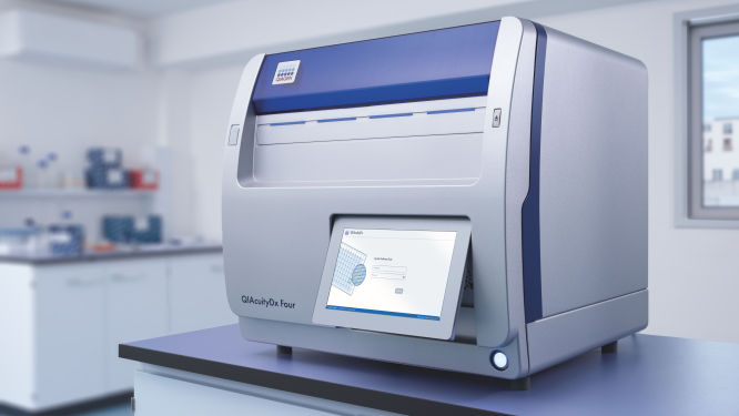
The QIAcuityDx dPCR System is intended for in vitro diagnostic use, using automated multiplex quantification dPCR technology, for the purpose of providing diagnostic information concerning pathological states.

QIAcuity and QIAcuityDx dPCR instruments are sold under license from Bio-Rad Laboratories, Inc. and exclude rights for use with pediatric applications. The QIAcuityDx medical device is currently under development and will be available in 20 countries in H2 2024.