


Digital PCR assays for colorectal cancer gene variants
Order ready-to-use dPCR assays
Revolutionizing colorectal cancer research with precision dPCR assays
The biological complexity and frequent late diagnoses of colorectal cancer pose significant challenges to researchers. Each case of colorectal cancer can vary significantly in terms of genetic mutations and clinical presentation, which necessitates precise characterization of gene variants to tailor effective treatments.
Digital PCR (dPCR) emerges as a vital tool in this context because it offers highly sensitive and accurate quantification of genetic material. This precision makes it possible to detect even low levels of genetic mutations that could be crucial for understanding the specific pathways involved in individual cases, thereby facilitating research into more targeted and effective therapies.
Explore colorectal cancer related dPCR assays by gene
A variety of gene variants are associated with the development and progression of colorectal cancer. Key mutations in genes such as APC, KRAS, TP53 and BRAF play significant roles in driving tumor growth and influencing resistance to therapies. Specifically, APC mutations are common in the early stages of carcinogenesis, marking it as an essential target for early detection. Meanwhile, KRAS mutations critically affect the tumor's responsiveness to certain chemotherapeutic agents and targeted therapies, underscoring the importance of personalized treatment plans.
Our collection of dPCR LNA Mutation Assays provides researchers with essential tools for in-depth analysis of these and other pivotal genetic changes. These assays facilitate precise quantification and characterization, contributing to the advancement of targeted research and therapeutic development.
Gene | Mutation Type | Mutation (CDS) | Mutation (AA) | COSMIC ID (COSV) | COSMIC ID (COSM) | Codon | dPCR Mutation Assay |
|---|---|---|---|---|---|---|---|
| BRAF | Substitution - Missense | c.1798G>A | p.V600M | COSV56075762 | COSM1130 | 600 | DMH0000218 |
| BRAF | Substitution - Missense | c.1798_1799delinsAA | p.V600K | COSV56057713 | COSM473 | 600 | DMH0000001 |
| BRAF | Substitution - Missense | c.1798_1799delinsAG | p.V600R | COSV56058419 | COSM474 | 600 | DMH0000002 |
| BRAF | Substitution - Missense | c.1799T>A | p.V600E | COSV56056643 | COSM476 | 600 | DMH0000004 |
| BRAF | Substitution - Missense | c.1799T>G | p.V600G | COSV56080151 | COSM6137 | 600 | DMH0000068 |
| BRAF | Substitution - Missense | c.1799_1800delinsAA | p.V600E | COSV56059110 | COSM475 | 600 | DMH0000003 |
| BRAF | Substitution - Missense | c.1799_1800delinsAT | p.V600D | COSV56059623 | COSM477 | 600 | DMH0000039 |
| EGFR | Substitution - Missense | c.2155G>A | p.G719S | COSV51767289 | COSM6252 | 719 | DMH0000055 |
| EGFR | Substitution - Missense | c.2155G>T | p.G719C | COSV51766606 | COSM6253 | 719 | DMH0000280 |
| EGFR | Substitution - Missense | c.2156G>C | p.G719A | COSV51769339 | COSM6239 | 719 | DMH0000057 |
| EGFR | Substitution - Missense | c.2303G>T | p.S768I | COSV51768106 | COSM6241 | 768 | DMH0000308 |
| EGFR | Substitution - Missense | c.2369C>T | p.T790M | COSV51765492 | COSM6240 | 790 | DMH0000085 |
| EGFR | Substitution - Missense | c.2573T>G | p.L858R | COSV51765161 | COSM6224 | 858 | DMH0000386 |
| EGFR | Substitution - Missense | c.2582T>A | p.L861Q | COSV51766344 | COSM6213 | 861 | DMH0000006 |
| KRAS | Substitution - Missense | c.34G>A | p.G12S | COSV55497461 | COSM517 | 12 | DMH0000519 |
| KRAS | Substitution - Missense | c.34G>C | p.G12R | COSV55497582 | COSM518 | 12 | DMH0000284 |
| KRAS | Substitution - Missense | c.34G>T | p.G12C | COSV55497469 | COSM516 | 12 | DMH0000309 |
| KRAS | Substitution - Missense | c.35G>A | p.G12D | COSV55497369 | COSM521 | 12 | DMH0000286 |
| KRAS | Substitution - Missense | c.35G>C | p.G12A | COSV55497479 | COSM522 | 12 | |
| KRAS | Substitution - Missense | c.35G>T | p.G12V | COSV55497419 | COSM520 | 12 | DMH0000285 |
| KRAS | Substitution - Missense | c.37G>A | p.G13S | COSV55509530 | COSM528 | 13 | DMH0000331 |
| KRAS | Substitution - Missense | c.37G>C | p.G13R | COSV55502117 | COSM529 | 13 | DMH0000332 |
| KRAS | Substitution - Missense | c.37G>T | p.G13C | COSV55497378 | COSM527 | 13 | DMH0000195 |
| KRAS | Substitution - Missense | c.38G>A | p.G13D | COSV55497388 | COSM532 | 13 | DMH0000289 |
| KRAS | Substitution - Missense | c.38G>C | p.G13A | COSV55497357 | COSM533 | 13 | DMH0000334 |
| KRAS | Substitution - Missense | c.38G>T | p.G13V | COSV55522580 | COSM534 | 13 | DMH0000527 |
| KRAS | Substitution - Missense | c.38_39delinsAT | p.G13D | COSV55508630 | COSM531 | 13 | DMH0000525 |
| KRAS | Substitution - Missense | c.181C>A | p.Q61K | COSV55502066 | COSM549 | 61 | DMH0000290 |
| KRAS | Substitution - Missense | c.181C>G | p.Q61E | COSV55502677 | COSM550 | 61 | DMH0000044 |
| KRAS | Substitution - Missense | c.182A>C | p.Q61P | COSV55508574 | COSM551 | 61 | DMH0000022 |
| KRAS | Substitution - Missense | c.182A>G | p.Q61R | COSV55498739 | COSM552 | 61 | DMH0000023 |
| KRAS | Substitution - Missense | c.182A>T | p.Q61L | COSV55504296 | COSM553 | 61 | DMH0000198 |
| KRAS | Substitution - Missense | c.183A>C | p.Q61H | COSV55498802 | COSM554 | 61 | DMH0000024 |
| KRAS | Substitution - Missense | c.183A>T | p.Q61H | COSV55499223 | COSM555 | 61 | DMH0000025 |
| NRAS | Substitution - Missense | c.34G>A | p.G12S | COSV54736621 | COSM563 | 12 | DMH0000188 |
| NRAS | Substitution - Missense | c.34G>C | p.G12R | COSV54736940 | COSM561 | 12 | DMH0000336 |
| NRAS | Substitution - Missense | c.34G>T | p.G12C | COSV54736487 | COSM562 | 12 | DMH0000186 |
| NRAS | Substitution - Missense | c.35G>C | p.G12A | COSV54736555 | COSM565 | 12 | DMH0000339 |
| NRAS | Substitution - Missense | c.35G>T | p.G12V | COSV54736974 | COSM566 | 12 | DMH0000340 |
| NRAS | Substitution - Missense | c.37G>A | p.G13S | COSV54736476 | COSM571 | 13 | DMH0000510 |
| NRAS | Substitution - Missense | c.37G>C | p.G13R | COSV54736550 | COSM569 | 13 | DMH0000341 |
| NRAS | Substitution - Missense | c.37G>T | p.G13C | COSV54736386 | COSM570 | 13 | DMH0000342 |
| NRAS | Substitution - Missense | c.38G>A | p.G13D | COSV54736416 | COSM573 | 13 | DMH0000343 |
| NRAS | Substitution - Missense | c.38G>C | p.G13A | COSV54736793 | COSM575 | 13 | DMH0000345 |
| NRAS | Substitution - Missense | c.38G>T | p.G13V | COSV54736480 | COSM574 | 13 | DMH0000344 |
| NRAS | Substitution - Missense | c.181C>A | p.Q61K | COSV54736310 | COSM580 | 61 | DMH0000505 |
| NRAS | Substitution - Missense | c.181C>G | p.Q61E | COSV54743343 | COSM581 | 61 | DMH0000347 |
| NRAS | Substitution - Missense | c.182A>G | p.Q61R | COSV54736340 | COSM584 | 61 | DMH0000183 |
| NRAS | Substitution - Missense | c.182A>T | p.Q61L | COSV54736624 | COSM583 | 61 | DMH0000190 |
| NRAS | Substitution - Missense | c.183A>C | p.Q61H | COSV54736320 | COSM586 | 61 | DMH0000180 |
| NRAS | Substitution - Missense | c.183A>T | p.Q61H | COSV54736991 | COSM585 | 61 | DMH0000349 |
| PIK3CA | Substitution - Missense | c.1633G>A | p.E545K | COSV55873239 | COSM763 | 545 | DMH0000292 |
| PIK3CA | Substitution - Missense | c.1634A>G | p.E545G | COSV55873220 | COSM764 | 545 | DMH0000033 |
| PIK3CA | Substitution - Missense | c.1636C>A | p.Q546K | COSV55873527 | COSM766 | 546 | DMH0000037 |
| PIK3CA | Substitution - Missense | c.1637A>G | p.Q546R | COSV55876869 | COSM12459 | 546 | DMH0000212 |
| PIK3CA | Substitution - Missense | c.3129G>T | p.M1043I | COSV55878974 | COSM773 | 1043 | DMH0000034 |
| PIK3CA | Substitution - Missense | c.3139C>T | p.H1047Y | COSV55876499 | COSM774 | 1047 | DMH0000209 |
| PIK3CA | Substitution - Missense | c.3140A>G | p.H1047R | COSV55873195 | COSM775 | 1047 | DMH0000036 |
| PIK3CA | Substitution - Missense | c.3140A>T | p.H1047L | COSV55873401 | COSM776 | 1047 | DMH0000062 |
| PIK3CA | Substitution - Missense | c.3145G>C | p.G1049R | COSV55874453 | COSM12597 | 1049 | DMH0000050 |
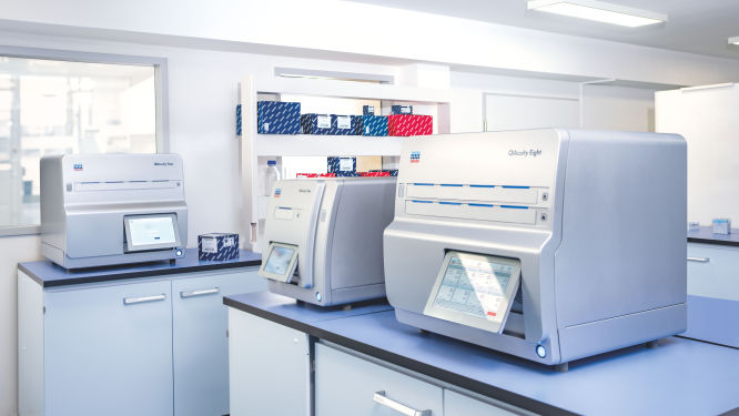
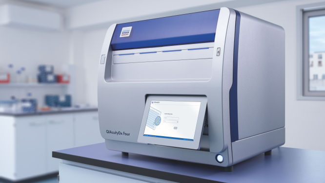
Discover the QIAcuity family of dPCR instruments
*FDA ‘Medical Devices; Laboratory Developed Tests’ final rule, May 6, 2024 and European Union regulation requirements on ‘In-House Assays’ (Regulation (EU) 2017/746 -IVDR- Art. 5(5))
Frequently asked questions
How do dPCR LNA Mutation Assays benefit cancer researchers?
How is KRAS involved in colorectal cancer?
What role does APC play in colorectal cancer?
How does MLH1 influence colorectal cancer?
What is the significance of MSH2 in colorectal cancer development?
How is MSH6 implicated in colorectal cancer mechanisms?
How is PIK3CA involved in colorectal cancer?
How does PMS2 involvement impact colorectal cancer?
What is the role of SMAD4 in the progression of colorectal cancer?
In what way is TP53 involved in colorectal cancer?
Disclaimers
dPCR LNA Mutation Assays are intended for molecular biology applications. These products are not intended for the diagnosis, prevention, or treatment of a disease.
The QIAcuity is intended for molecular biology applications. This product is not intended for the diagnosis, prevention or treatment of a disease. Therefore, the performance characteristics of the product for clinical use (i.e., diagnostic, prognostic, therapeutic or blood banking) is unknown.
The QIAcuityDx dPCR System is intended for in vitro diagnostic use, using automated multiplex quantification dPCR technology, for the purpose of providing diagnostic information concerning pathological states.
QIAcuity and QIAcuityDx dPCR instruments are sold under license from Bio-Rad Laboratories, Inc. and exclude rights for use with pediatric applications. The QIAcuityDx medical device is currently under development and will be available in 20 countries in H2 2024.