


Digital PCR assays for lung cancer gene variants
Order ready-to-use dPCR assays
Revolutionizing lung cancer research with precision dPCR assays
Lung cancer presents significant challenges to oncology researchers due to its genetic complexity and mutation diversity. The development of personalized therapies relies on accurately detecting and understanding these gene variants. Digital PCR (dPCR) technology, known for its precision and sensitivity, can play a pivotal role in identifying these markers and advancing research, in particular where precise identification of low prevalence biomarkers is required.
Our leading portfolio of dPCR LNA Mutation Assays offers unmatched accuracy, sensitivity and reproducibility, enabling precise detection and quantification of key gene variants. These capabilities are essential for advancing targeted research and therapies, facilitating the development of more personalized treatment approaches.
Explore lung cancer related dPCR assays by gene
Lung cancer's complexity is underscored by its subtypes, each characterized by unique genetic markers and treatment responses. The most common subtype, non-small cell lung cancer (NSCLC), represents about 85% of cases and includes adenocarcinoma, squamous cell carcinoma and large cell carcinoma. Small cell lung cancer (SCLC) comprises most of the remaining cases.
Specific mutations in genes such as EGFR, KRAS, BRAF and others are pivotal in advancing our understanding of lung cancer progression and identifying potential therapeutic targets. These genetic variations also play a crucial role in studying the mechanisms of treatment resistance, informing the development of more effective targeted therapies. Our assay collection provides a robust toolkit for advanced oncology research, facilitating precise genetic analysis and insights.
Gene | Mutation Type | Mutation (CDS) | Mutation (AA) | COSMIC ID (COSV) | COSMIC ID (COSM) | Codon | dPCR Mutation Assay |
|---|---|---|---|---|---|---|---|
| BRAF | Substitution - Missense | c.1406G>C | p.G469A | COSV56061424 | COSM460 | 469 | DMH0000047 |
| BRAF | Substitution - Missense | c.1798G>A | p.V600M | COSV56075762 | COSM1130 | 600 | DMH0000218 |
| BRAF | Substitution - Missense | c.1798_1799delinsAA | p.V600K | COSV56057713 | COSM473 | 600 | DMH0000001 |
| BRAF | Substitution - Missense | c.1798_1799delinsAG | p.V600R | COSV56058419 | COSM474 | 600 | DMH0000002 |
| BRAF | Substitution - Missense | c.1799T>A | p.V600E | COSV56056643 | COSM476 | 600 | DMH0000004 |
| BRAF | Substitution - Missense | c.1799T>G | p.V600G | COSV56080151 | COSM6137 | 600 | DMH0000068 |
| BRAF | Substitution - Missense | c.1799_1800delinsAA | p.V600E | COSV56059110 | COSM475 | 600 | DMH0000003 |
| BRAF | Substitution - Missense | c.1799_1800delinsAT | p.V600D | COSV56059623 | COSM477 | 600 | DMH0000039 |
| EGFR | Substitution - Missense | c.2125G>A | p.E709K | COSV51767260 | COSM12988 | 709 | DMH0000041 |
| EGFR | Substitution - Missense | c.2126A>C | p.E709A | COSV51772956 | COSM13427 | 709 | DMH0000271 |
| EGFR | Substitution - Missense | c.2126A>T | p.E709V | COSV51766584 | COSM12371 | 709 | DMH0000397 |
| EGFR | Substitution - Missense | c.2155G>A | p.G719S | COSV51767289 | COSM6252 | 719 | DMH0000055 |
| EGFR | Substitution - Missense | c.2155G>T | p.G719C | COSV51766606 | COSM6253 | 719 | DMH0000280 |
| EGFR | Substitution - Missense | c.2156G>C | p.G719A | COSV51769339 | COSM6239 | 719 | DMH0000057 |
| EGFR | c.2233_2247del | p.K745_E749del | COSV51769442 | COSM26038 | 745-749 | DMH0000082 | |
| EGFR | c.2235_2249del | p.E746_A750del | COSV51765119 | COSM6223 | 746-750 | DMH0000276 | |
| EGFR | c.2236_2250del | p.E746_A750del | COSV51765066 | COSM6225 | 746-750 | DMH0000240 | |
| EGFR | c.2236_2253del | p.E746_T751del | COSV51811139 | COSM12728 | 746-751 | DMH0000250 | |
| EGFR | c.2237_2251del | p.E746_T751delinsA | COSV51769364 | COSM12678 | 746-751 | DMH0000258 | |
| EGFR | c.2237_2254del | p.E746_S752delinsA | COSV51769704 | COSM12367 | 746-752 | DMH0000072 | |
| EGFR | c.2238_2255del | p.E746_S752delinsD | COSV51772418 | COSM6220 | 746-752 | DMH0000232 | |
| EGFR | c.2239_2247del | p.L747_E749del | COSV51780076 | COSM6218 | 747-749 | DMH0000262 | |
| EGFR | c.2239_2256del | p.L747_S752del | COSV51767308 | COSM6255 | 747-752 | DMH0000076 | |
| EGFR | c.2240_2254del | p.L747_T751del | COSV51766247 | COSM12369 | 747-751 | DMH0000275 | |
| EGFR | c.2240_2257del | p.L747_P753delinsS | COSV51767961 | COSM12370 | 747-753 | DMH0000266 | |
| EGFR | c.2253_2276del | p.S752_I759del | COSV51766231 | COSM13556 | 752-759 | DMH0000078 | |
| EGFR | Substitution - Missense | c.2303G>T | p.S768I | COSV51768106 | COSM6241 | 768 | DMH0000308 |
| EGFR | c.2310_2311insGGT | p.D770_N771insG | COSV51769298 | COSM12378 | 770-771 | DMH0000248 | |
| EGFR | Substitution - Missense | c.2369C>T | p.T790M | COSV51765492 | COSM6240 | 790 | DMH0000085 |
| EGFR | Substitution - Missense | c.2389T>A | p.C797S | COSV51766493 | COSM6493937 | 797 | DMH0000007 |
| EGFR | Substitution - Missense | c.2390G>C | p.C797S | COSV51766509 | COSM5945664 | 797 | DMH0000052 |
| EGFR | Substitution - Missense | c.2573T>G | p.L858R | COSV51765161 | COSM6224 | 858 | DMH0000386 |
| EGFR | Substitution - Missense | c.2582T>A | p.L861Q | COSV51766344 | COSM6213 | 861 | DMH0000006 |
| KRAS | Substitution - Missense | c.34G>A | p.G12S | COSV55497461 | COSM517 | 12 | DMH0000519 |
| KRAS | Substitution - Missense | c.34G>C | p.G12R | COSV55497582 | COSM518 | 12 | DMH0000284 |
| KRAS | Substitution - Missense | c.34G>T | p.G12C | COSV55497469 | COSM516 | 12 | DMH0000309 |
| KRAS | Substitution - Missense | c.35G>A | p.G12D | COSV55497369 | COSM521 | 12 | DMH0000286 |
| KRAS | Substitution - Missense | c.35G>C | p.G12A | COSV55497479 | COSM522 | 12 | |
| KRAS | Substitution - Missense | c.35G>T | p.G12V | COSV55497419 | COSM520 | 12 | DMH0000285 |
| KRAS | Substitution - Missense | c.37G>A | p.G13S | COSV55509530 | COSM528 | 13 | DMH0000331 |
| KRAS | Substitution - Missense | c.37G>C | p.G13R | COSV55502117 | COSM529 | 13 | DMH0000332 |
| KRAS | Substitution - Missense | c.37G>T | p.G13C | COSV55497378 | COSM527 | 13 | DMH0000195 |
| KRAS | Substitution - Missense | c.38G>A | p.G13D | COSV55497388 | COSM532 | 13 | DMH0000289 |
| KRAS | Substitution - Missense | c.38G>C | p.G13A | COSV55497357 | COSM533 | 13 | DMH0000334 |
| KRAS | Substitution - Missense | c.38G>T | p.G13V | COSV55522580 | COSM534 | 13 | DMH0000527 |
| KRAS | Substitution - Missense | c.38_39delinsAT | p.G13D | COSV55508630 | COSM531 | 13 | DMH0000525 |
| KRAS | Substitution - Missense | c.181C>A | p.Q61K | COSV55502066 | COSM549 | 61 | DMH0000290 |
| KRAS | Substitution - Missense | c.181C>G | p.Q61E | COSV55502677 | COSM550 | 61 | DMH0000044 |
| KRAS | Substitution - Missense | c.182A>C | p.Q61P | COSV55508574 | COSM551 | 61 | DMH0000022 |
| KRAS | Substitution - Missense | c.182A>G | p.Q61R | COSV55498739 | COSM552 | 61 | DMH0000023 |
| KRAS | Substitution - Missense | c.182A>T | p.Q61L | COSV55504296 | COSM553 | 61 | DMH0000198 |
| KRAS | Substitution - Missense | c.183A>C | p.Q61H | COSV55498802 | COSM554 | 61 | DMH0000024 |
| KRAS | Substitution - Missense | c.183A>T | p.Q61H | COSV55499223 | COSM555 | 61 | DMH0000025 |
| NRAS | Substitution - Missense | c.34G>A | p.G12S | COSV54736621 | COSM563 | 12 | DMH0000188 |
| NRAS | Substitution - Missense | c.34G>C | p.G12R | COSV54736940 | COSM561 | 12 | DMH0000336 |
| NRAS | Substitution - Missense | c.34G>T | p.G12C | COSV54736487 | COSM562 | 12 | DMH0000186 |
| NRAS | Substitution - Missense | c.35G>C | p.G12A | COSV54736555 | COSM565 | 12 | DMH0000339 |
| NRAS | Substitution - Missense | c.35G>T | p.G12V | COSV54736974 | COSM566 | 12 | DMH0000340 |
| NRAS | Substitution - Missense | c.37G>A | p.G13S | COSV54736476 | COSM571 | 13 | DMH0000510 |
| NRAS | Substitution - Missense | c.37G>C | p.G13R | COSV54736550 | COSM569 | 13 | DMH0000341 |
| NRAS | Substitution - Missense | c.37G>T | p.G13C | COSV54736386 | COSM570 | 13 | DMH0000342 |
| NRAS | Substitution - Missense | c.38G>A | p.G13D | COSV54736416 | COSM573 | 13 | DMH0000343 |
| NRAS | Substitution - Missense | c.38G>C | p.G13A | COSV54736793 | COSM575 | 13 | DMH0000345 |
| NRAS | Substitution - Missense | c.38G>T | p.G13V | COSV54736480 | COSM574 | 13 | DMH0000344 |
| NRAS | Substitution - Missense | c.181C>A | p.Q61K | COSV54736310 | COSM580 | 61 | DMH0000505 |
| NRAS | Substitution - Missense | c.181C>G | p.Q61E | COSV54743343 | COSM581 | 61 | DMH0000347 |
| NRAS | Substitution - Missense | c.182A>G | p.Q61R | COSV54736340 | COSM584 | 61 | DMH0000183 |
| NRAS | Substitution - Missense | c.182A>T | p.Q61L | COSV54736624 | COSM583 | 61 | DMH0000190 |
| NRAS | Substitution - Missense | c.183A>C | p.Q61H | COSV54736320 | COSM586 | 61 | DMH0000180 |
| NRAS | Substitution - Missense | c.183A>T | p.Q61H | COSV54736991 | COSM585 | 61 | DMH0000349 |
| PIK3CA | Substitution - Missense | c.1633G>A | p.E545K | COSV55873239 | COSM763 | 545 | DMH0000292 |
| PIK3CA | Substitution - Missense | c.1634A>G | p.E545G | COSV55873220 | COSM764 | 545 | DMH0000033 |
| PIK3CA | Substitution - Missense | c.3129G>T | p.M1043I | COSV55878974 | COSM773 | 1043 | DMH0000034 |
| PIK3CA | Substitution - Missense | c.3139C>T | p.H1047Y | COSV55876499 | COSM774 | 1047 | DMH0000209 |
| PIK3CA | Substitution - Missense | c.3140A>G | p.H1047R | COSV55873195 | COSM775 | 1047 | DMH0000036 |
| PIK3CA | Substitution - Missense | c.3140A>T | p.H1047L | COSV55873401 | COSM776 | 1047 | DMH0000062 |
| PTEN | Substitution - Missense | c.388C>G | p.R130G | COSV64288384 | COSM5219 | 130 | DMH0000296 |
| PTEN | Substitution - Nonsense | c.388C>T | p.R130* | COSV64288463 | COSM5152 | 130 | DMH0000294 |
| PTEN | Substitution - Nonsense | c.697C>T | p.R233* | COSV64288653 | COSM5154 | 233 | DMH0000295 |
| TP53 | Substitution - Missense | c.488A>G | p.Y163C | COSV52663142 | COSM10808 | 163 | DMH0000112 |
| TP53 | Substitution - Missense | c.517G>T | p.V173L | COSV52676535 | COSM43559 | 173 | DMH0000126 |
| TP53 | Substitution - Missense | c.578A>G | p.H193R | COSV52662414 | COSM10742 | 193 | DMH0000108 |
| TP53 | Substitution - Missense | c.659A>G | p.Y220C | COSV52661282 | COSM10758 | 220 | DMH0000440 |
| TP53 | Substitution - Missense | c.743G>T | p.R248L | COSV52675468 | COSM6549 | 248 | DMH0000381 |
| TP53 | Substitution - Missense | c.818G>A | p.R273H | COSV52660980 | COSM10660 | 273 | DMH0000094 |
| TP53 | c.818G>T | p.R273L | COSV52664805 | COSM10779 | 273 | DMH0000114 | |
| TP53 | c.856G>A | p.E286K | COSV52664318 | COSM10726 | 286 | DMH0000364 |
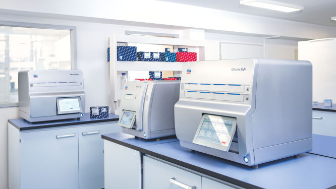
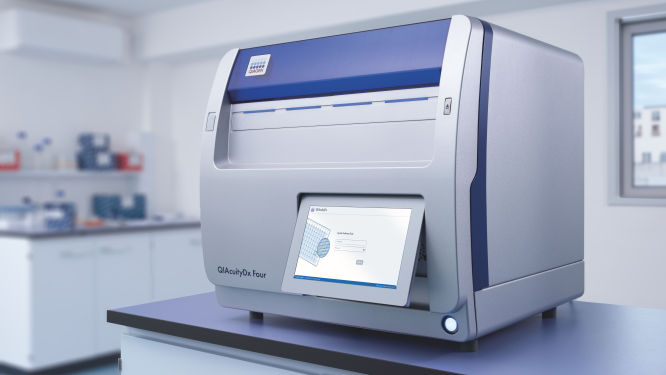
Discover the QIAcuity family of dPCR instruments
*FDA ‘Medical Devices; Laboratory Developed Tests’ final rule, May 6, 2024 and European Union regulation requirements on ‘In-House Assays’ (Regulation (EU) 2017/746 -IVDR- Art. 5(5))
Frequently asked questions
How do dPCR LNA Mutation Assays benefit cancer researchers?
What role does EGFR play in lung cancer?
How is KRAS involved in lung cancer?
How does BRAF mutation affect lung cancer?
What is the significance of ALK in lung cancer?
How does TP53 contribute to lung cancer?
What is the role of MET in lung cancer?
In what way is PIK3CA associated with lung cancer?
How are ROS1 rearrangements important in lung cancer?
What impact do RET rearrangements have on lung cancer?
How does the loss of RB1 function affect lung cancer?
Disclaimers
dPCR LNA Mutation Assays are intended for molecular biology applications. These products are not intended for the diagnosis, prevention, or treatment of a disease.
The QIAcuity is intended for molecular biology applications. This product is not intended for the diagnosis, prevention or treatment of a disease. Therefore, the performance characteristics of the product for clinical use (i.e., diagnostic, prognostic, therapeutic or blood banking) is unknown.
The QIAcuityDx dPCR System is intended for in vitro diagnostic use, using automated multiplex quantification dPCR technology, for the purpose of providing diagnostic information concerning pathological states.
QIAcuity and QIAcuityDx dPCR instruments are sold under license from Bio-Rad Laboratories, Inc. and exclude rights for use with pediatric applications. The QIAcuityDx medical device is currently under development and will be available in 20 countries in H2 2024.