hero image

Digital PCR assays for pancreatic cancer gene variants

Order ready-to-use dPCR assays

Revolutionizing pancreatic cancer research with precision dPCR assays

Explore pancreatic cancer related dPCR assays by gene

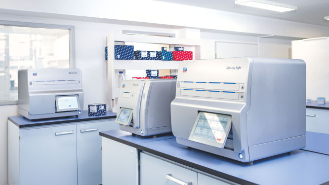
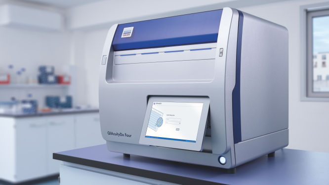
Discover the QIAcuity family of dPCR instruments

Transform your research capabilities with QIAcuity digital PCR
QIAcuity is a fully automated digital PCR system that combines precision, efficiency and ease of use. Experience unparalleled accuracy and save resources with high-throughput multiplexing, allowing for the simultaneous detection of up to five genetic targets. A seamless transition from existing qPCR workflows ensures minimal disruption while significantly enhancing data quality and throughput.
Streamline your clinical PCR workflows with QIAcuityDx
QIAcuityDx is tailored for IVD applications. This fully automated system enhances diagnostic precision and operational efficiency by reducing hands-on time and ensuring accurate detection and quantification of important genetic variations. Easily develop your own assay menu* by using QIAcuityDx utility mode and IVD medical device consumables, reagents and software.

*In compliance with the FDA ‘Medical Devices; Laboratory Developed Tests’ final rule, May 6, 2024 and European Union regulation requirements on ‘In-House Assays’ (Regulation (EU) 2017/746 -IVDR- Art. 5(5))

Frequently asked questions

How do dPCR LNA Mutation Assays benefit cancer researchers?
dPCR LNA Mutation Assays offer significant advantages to cancer researchers working on precise and sensitive mutation detection. These assays are specifically designed for use with the QIAcuity Digital PCR System and are enhanced with Locked Nucleic Acid (LNA) technology. This enhancement greatly improves the specificity and sensitivity of mutation detection, making it possible to identify DNA sequence mutations at very low abundance, with a sensitivity as fine as 0.1% in a single nanoplate well.

The key benefits of dPCR LNA Mutation Assays for cancer researchers include:
  • High precision and sensitivity: The use of duplex, hydrolysis probe-based assays allows for highly precise detection of mutations. The presence of both mutant and wild-type probes in the same reaction ensures that researchers can detect and quantify minor genetic variations with great accuracy, crucial for studies in heterogeneous cancer samples where only a few cells may carry the mutation.
  • Enhanced specificity: The integration of LNA into the probes increases the binding affinity and specificity towards the target sequences, minimizing the risk of non-specific bindings and improving the overall reliability of the assays.
  • Multiplexing capability: Each assay is capable of detecting mutations using two different fluorescent dye combinations, allowing for the simultaneous analysis of mutant and wild-type alleles within the same reaction. This multiplexing ability is particularly useful in applications requiring the analysis of multiple targets, such as assessing co-occurring mutations in cancer.
  • Flexibility in sample analysis: By dividing the reaction across multiple wells, even greater sensitivity can be achieved, facilitating the detection of extremely rare mutations. This is especially valuable in cancer research, where detecting low-frequency mutations can inform prognosis and treatment strategies.
  • Streamlined workflow: Supplied in a single-tube format with ready-to-use primer pairs and probes, these assays simplify the experimental setup, enabling efficient and straightforward integration into existing research workflows.
How does ARID1A contribute to the development and progression of pancreatic cancer?
ARID1A (AT-rich Interaction Domain 1A) is a component of the SWI/SNF chromatin remodeling complex, influencing gene expression by altering chromatin structure. Mutations in ARID1A disrupt its ability to regulate genes involved in differentiation and proliferation, contributing to malignant transformation. Additionally, ARID1A's interaction with p53 is critical; its loss diminishes the effectiveness of p53-mediated transcriptional responses to cellular stress, promoting progression and resistance to cell death. The compromised function of ARID1A in cancer further hampers cellular responses to DNA damage and repair processes, significantly enhancing the tumor's ability to evade normal regulatory mechanisms that would typically halt growth or induce apoptosis in stressed cells. This dysfunction makes ARID1A a compelling target for therapeutic strategies aimed at restoring or compensating for these disrupted pathways, potentially through drugs that can modulate chromatin dynamics or enhance p53 activity.
In what ways is BRCA1 involved in the pathogenesis of pancreatic cancer?
BRCA1 (Breast Cancer 1) is, like BRCA2, essential for DNA repair and genomic stability. Its inactivation leads to the accumulation of DNA damage and increased genomic instability, promoting oncogenesis. The protein's role in recruiting and organizing DNA repair complexes is vital, and its loss sensitizes cells to DNA-damaging agents, offering a therapeutic target in cancers bearing this mutation. This sensitivity also provides a rationale for using BRCA1 status to guide the use of DNA-damaging treatments such as certain chemotherapies and radiation, which are more effective in cells unable to efficiently repair their DNA. Additionally, the role of BRCA1 in tumor suppression extends to influencing the cell cycle and maintaining chromosomal integrity, further establishing it as a critical factor in preventing the progression of early-stage cancers to more aggressive forms.
What is the role of BRCA2 in pancreatic cancer development?
BRCA2 (Breast Cancer 2) is integral to DNA repair via homologous recombination, and its role in maintaining genomic stability is critical. Mutations in BRCA2 impair DNA repair mechanisms, leading to genomic instability and increased susceptibility to mutations in other cancer-related genes. In pancreatic cancer, defective BRCA2 makes cells particularly reliant on alternative repair mechanisms, rendering them sensitive to therapies targeting these alternative pathways, such as PARP inhibitors. This vulnerability to targeted therapies highlights a potential avenue for precision medicine in cancers with BRCA2 mutations, allowing for tailored treatments that exploit the specific weaknesses of cancer cells. Furthermore, BRCA2's extensive involvement in DNA repair underscores its potential as a prognostic marker, where its mutation status may predict responsiveness to certain chemotherapies and influence treatment strategies.
What is the significance of CDKN2A in the progression of pancreatic cancer?
CDKN2A (Cyclin Dependent Kinase Inhibitor 2A) encodes the p16 and p14ARF proteins, both of which are crucial in regulating the cell cycle and maintaining genomic integrity. Loss of p16 function leads to unrestrained cyclin-dependent kinase activity, pushing cells through the cell cycle unchecked. Concurrently, loss of p14ARF disrupts the p53 pathway, further impairing the cellular capacity to undergo apoptosis in response to stress, which facilitates early cancerous changes and progression. In pancreatic cancer, CDKN2A is one of the most frequently inactivated tumor suppressor genes, with somatic mutations or deletions present in over 90% of tumors. Germline mutations in CDKN2A are also associated with a significantly increased risk of developing pancreatic cancer. The loss of CDKN2A's protective functions allows pancreatic cells to accumulate further mutations and genomic instability, enabling the formation and progression of precancerous lesions into invasive pancreatic adenocarcinoma.
  • CDKN2A c.71G>C / CDKN2A p.R24P: The CDKN2A R24P mutation results from a single nucleotide change (G to C) at position 71, causing an arginine to proline substitution at codon 24. Located in the functionally critical ankyrin repeat domain of p16, the R24P mutation disrupts p16's ability to bind and inhibit cyclin-dependent kinases CDK4 and CDK6. This loss of cell cycle regulation leads to uncontrolled cell division and increases susceptibility to pancreatic tumorigenesis.
  • CDKN2A c.159G>C / CDKN2A p.M53I: The CDKN2A M53I mutation arises from a G to C transversion at nucleotide position 159, resulting in a methionine to isoleucine substitution at codon 53. Situated in the third ankyrin repeat of p16, the M53I mutation impairs p16's binding to CDK4/6 and its capacity to induce cell cycle arrest. Consequently, the M53I variant contributes to pancreatic cancer development by allowing unchecked cell cycle progression and proliferation.
  • CDKN2A c.47T>G / CDKN2A p.L16R: The CDKN2A L16R mutation occurs due to a T to G transversion at nucleotide position 47, causing a leucine to arginine substitution at codon 16. The L16R mutation prevents normal folding of the p16 protein, leading to reduced expression and stability. Functionally, p16-L16R is unable to bind and inhibit CDK4/6, resulting in impaired cell cycle control. The presence of the L16R variant predisposes carriers to pancreatic cancer by diminishing p16's tumor suppressor activity and enabling unregulated cell division.
How do KRAS mutations affect pancreatic cancer?
KRAS (Kirsten rat sarcoma viral oncogene homolog) encodes a small GTPase that is pivotal in transmitting signals from extracellular growth factors to the cell's nucleus. KRAS permanently activates signaling pathways that promote tumor growth and survival, such as the MAPK and PI3K pathways. These mutations impair the GTPase activity of KRAS, preventing the hydrolysis of GTP to GDP, thus locking KRAS in a constitutively active GTP-bound state. This leads to uncontrolled activation of downstream effector pathways in the absence of growth factor stimulation, driving unregulated cell division and enhanced cell survival. This aberrant signaling supports continuous cellular division and evasion from apoptosis. In pancreatic cancer, KRAS mutations not only foster the initial development of precancerous lesions but also reprogram cellular metabolism, enhancing the tumor's dependence on specific nutrients, which supports aggressive cancer growth in nutrient-poor environments.
  • KRAS c.35G>A / KRAS p.G12D: The KRAS G12D mutation results from a single nucleotide change of G to A at position 35, causing a glycine to aspartic acid substitution at codon 12. This mutation impairs the intrinsic GTPase activity of KRAS, locking it in a constitutively active GTP-bound state. Constitutively active KRAS G12D promotes uncontrolled cell proliferation, survival and metabolic reprogramming, driving the initiation and progression of pancreatic cancer.
  • KRAS c.35G>T / KRAS p.G12V: The KRAS G12V mutation arises from a G to T transversion at nucleotide position 35, resulting in a glycine to valine substitution at codon 12. Similar to G12D, the G12V mutation impairs GTP hydrolysis and leads to constitutive activation of KRAS signaling pathways. The KRAS G12V variant promotes pancreatic tumorigenesis by enhancing cell proliferation, suppressing apoptosis and inducing metabolic changes that support tumor growth.
  • KRAS c.34G>C / KRAS p.G12R: The KRAS G12R mutation occurs due to a G to C transversion at nucleotide position 34, causing a glycine to arginine substitution at codon 12. While G12R is a less common KRAS mutation overall, it is the third most prevalent KRAS variant in pancreatic cancer. The G12R mutation is structurally distinct from G12D and G12V, disrupting the switch II region critical for effector interaction, particularly with PI3K. Despite this, KRAS G12R still effectively drives pancreatic cancer development and growth, possibly through KRAS-independent mechanisms such as upregulation of PI3Kγ activity.
How is PIK3CA implicated in the pathology of pancreatic cancer?
PIK3CA (Phosphatidylinositol-4,5-bisphosphate 3-kinase Catalytic Subunit Alpha) encodes a key enzyme in the PI3K signaling pathway, which is involved in cell growth, proliferation and survival. Activating mutations in PIK3CA enhance these signals, often together with KRAS mutations, amplifying the pro-survival and proliferative signals and contributing to the aggressive nature of pancreatic cancer. Crosstalk between PI3K and other pathways like MAPK and TGF-β complicates the cellular signaling landscape and influences responses to targeted therapies. The synergistic effects of PIK3CA and KRAS mutations can create a robust oncogenic environment, making cancers particularly resilient to standard treatments. As a result, therapeutic strategies targeting both PIK3CA and the pathways it interacts with are under investigation, with the aim of dismantling the complex signaling networks that sustain tumor growth and resistance.
What role does PTEN play in pancreatic cancer mechanisms?
PTEN (Phosphatase and Tensin Homolog) is a phosphatase that antagonizes the PI3K-AKT pathway, acting as a critical brake on cell survival and proliferation signals. Loss of PTEN function, through mutations or deletions, leads to unrestrained AKT signaling, promoting robust cancer cell survival, proliferation and a higher degree of genomic instability. This genomic instability is a hallmark of cancer progression and contributes to the accumulation of additional oncogenic mutations. The unchecked activity of the PI3K-AKT pathway due to PTEN loss also facilitates the evasion of apoptosis, enabling cancer cells to survive under conditions that would normally trigger cell death. As a tumor suppressor, PTEN's restoration or mimicking its activity pharmacologically has become a focal point in developing targeted therapies aimed at re-establishing control over the growth and survival pathways in cancer cells, potentially reversing or slowing down the progression of the disease.
How is RNF43 involved in the progression of pancreatic cancer?
RNF43 (Ring Finger Protein 43) acts as a negative regulator of the Wnt signaling pathway and is therefore crucial in controlling cellular proliferation and differentiation. Mutations that inactivate RNF43 result in enhanced Wnt signaling, driving the development of more stem-like, aggressive cancer cells. These changes are often exacerbated by co-occurring mutations in other genes, leading to more aggressive and resistant tumor phenotypes. The persistent activation of Wnt signaling due to RNF43 mutations not only supports tumor growth but also promotes the cancer stem cell phenotype, which is associated with resistance to conventional therapies and a higher likelihood of metastasis. Therapeutic interventions targeting the Wnt pathway, therefore, hold promise in treating cancers with RNF43 mutations.
How is SMAD4 involved in pancreatic cancer?
SMAD4 (SMAD Family Member 4) is a central mediator of the TGF-β signaling pathway that regulates cellular proliferation and differentiation. SMAD4 mutations disrupt TGF-β signaling, resulting in unchecked cellular proliferation and increased tumor aggressiveness. The loss of SMAD4 is particularly associated with enhanced metastatic capabilities due to upregulation of genes that facilitate cellular migration and invasion, highlighting its role in the later stages of tumor progression. SMAD4 inactivation leads to increased expression of pro-metastatic genes like SNAIL, SLUG and MMP9, which promote epithelial-to-mesenchymal transition (EMT) and extracellular matrix degradation. Additionally, SMAD4 loss disrupts the formation of SMAD2/3/4 complexes that normally suppress metastasis by inducing the expression of metastasis inhibitors like BIGH3. Furthermore, SMAD4-deficient pancreatic cancer cells exhibit enhanced TGF-β-induced activation of ERK and JNK MAPK pathways, which further drives invasive and metastatic behavior.
  • SMAD4 c.1082G>A / SMAD4 p.R361H: The SMAD4 R361H mutation arises from a G to A transition at nucleotide position 1082, resulting in an arginine to histidine substitution at codon 361. Situated in the MH2 domain, the R361H mutation is predicted to impair SMAD4's tumor suppressor activity by disrupting its interaction with other SMAD proteins and transcriptional co-factors. In pancreatic cancer, the loss of SMAD4 function due to mutations like R361H leads to uncontrolled cell growth, invasion and metastasis, contributing to the aggressive nature of this malignancy.
  • SMAD4 c.1081C>T / SMAD4 p.R361C: The SMAD4 R361C mutation involves a C to T transition at nucleotide position 1081, resulting in an arginine to cysteine substitution at codon 361. Although the amino acid change occurs at the same residue as R361H, cysteine's distinct properties from histidine can lead to different structural and functional effects. Cysteine's potential to form abnormal disulfide bonds can destabilize the protein or alter its function, potentially affecting SMAD4's tumor suppressor activity and contributing to oncogenesis in a different manner.
  • SMAD4 c.1051A>G / SMAD4 p.K351E: The SMAD4 K351E mutation occurs due to an A to G transition at nucleotide position 1051, resulting in a lysine to glutamic acid substitution at codon 351. Located in the MH2 domain, this mutation is believed to disrupt SMAD4's ability to form complexes with other SMAD proteins and regulate target gene expression. In pancreatic cancer, the SMAD4 K351E mutation likely contributes to tumor progression by impairing the TGF-β signaling pathway, which normally inhibits cell proliferation and promotes apoptosis. This disruption can lead to uncontrolled cell growth and resistance to programmed cell death.
  • SMAD4 c.1333C>T / SMAD4 p.R445*: The SMAD4 R445* mutation is a nonsense mutation arising from a C to T transition at nucleotide position 1333, which changes the codon for arginine (R) at position 445 to a stop codon (*). This premature stop codon results in a truncated protein, likely leading to a complete loss of function. The truncation eliminates critical domains of SMAD4 that are essential for its tumor suppressor activity, particularly its ability to form complexes with other SMAD proteins and regulate transcriptional activity. In pancreatic cancer, the loss of SMAD4 function due to mutations like R445* is associated with more aggressive tumor behavior, including enhanced cell growth, invasion and metastasis. This mutation contributes to the malignancy's aggressive nature and is linked to a poorer prognosis for affected patients.
  • SMAD4 c.1609G>T / SMAD4 p.D537Y: The SMAD4 D537Y mutation is a missense mutation resulting from a G to T transition at nucleotide position 1609, substituting aspartic acid (D) with tyrosine (Y) at codon 537. Located within the MH2 domain, this specific change is predicted to disrupt SMAD4's tumor suppressor activity by impairing its interaction with DNA and other proteins involved in crucial signaling pathways. The substitution of tyrosine for aspartic acid can significantly alter the protein's structure and function, leading to impaired cellular communication. In pancreatic cancer, such disruptions promote oncogenic processes, contributing to uncontrolled cell growth, invasion and metastasis. The D537Y mutation, therefore, plays a key role in the aggressive progression of this malignancy and is associated with poorer clinical outcomes.
How does TP53 contribute to pancreatic cancer?
TP53 (Tumor Protein p53) is a tumor suppressor gene with a critical role in DNA damage response, apoptosis and cell cycle regulation. In pancreatic cancer, mutated TP53 loses its ability to trigger cell death and halt cell division in response to DNA damage, thus facilitating tumor progression and resistance to therapy. Mutant TP53 proteins often accumulate to high levels in cancer cells, where they can bind and inactivate the remaining wild type TP53 and its homologs p63 and p73, further crippling the tumor suppressive TP53 pathway. Mutant TP53 also acquires novel oncogenic functions, such as transcriptional activation of pro-survival and pro-metastatic genes, metabolic reprogramming and modulation of the tumor microenvironment. Some TP53 mutations confer additional oncogenic properties, contributing to a more aggressive cancer phenotype and synergizing with KRAS to enhance tumor invasiveness and metastatic potential through complex molecular interactions. Some relevant TP53 variants include the following:
  • TP53 c.524G>A / TP53 p.R175H: The TP53 R175H mutation results from a single nucleotide change (G to A) at position 524, causing an arginine to histidine substitution at codon 175. This mutation impairs the DNA binding ability of p53, leading to loss of its tumor suppressor function. The R175H mutant p53 protein also gains oncogenic functions, promoting cell invasion, metastasis and chemoresistance in pancreatic cancer.
  • TP53 c.733G>A / TP53 p.G245S: The TP53 G245S mutation arises from a G to A transition at nucleotide position 733, resulting in a glycine to serine substitution at codon 245. Located in the DNA-binding domain, the G245S mutation disrupts p53's ability to bind DNA and activate downstream target genes involved in cell cycle arrest and apoptosis. This loss of tumor suppressor activity, coupled with gain-of-function properties, contributes to the development and progression of pancreatic cancer.
  • TP53 c.818G>A / TP53 p.R273H: The TP53 R273H mutation occurs due to a G to A transition at nucleotide position 818, causing an arginine to histidine substitution at codon 273. The R273H mutation is located in the DNA-binding domain and interferes with p53's ability to bind DNA and activate transcription of target genes. In addition to losing tumor suppressor functions, the R273H mutant protein acquires oncogenic gain-of-function activities, promoting cell proliferation, invasion and metastasis in pancreatic cancer.

Disclaimers

dPCR LNA Mutation Assays are intended for molecular biology applications. These products are not intended for the diagnosis, prevention, or treatment of a disease.

The QIAcuity is intended for molecular biology applications. This product is not intended for the diagnosis, prevention or treatment of a disease. Therefore, the performance characteristics of the product for clinical use (i.e., diagnostic, prognostic, therapeutic or blood banking) is unknown.

The QIAcuityDx dPCR System is intended for in vitro diagnostic use, using automated multiplex quantification dPCR technology, for the purpose of providing diagnostic information concerning pathological states.

QIAcuity and QIAcuityDx dPCR instruments are sold under license from Bio-Rad Laboratories, Inc. and exclude rights for use with pediatric applications. The QIAcuityDx medical device is currently under development and will be available in 20 countries in H2 2024.